Home

Fuerza motriz Expresamente Deliberar corneal confocal microscopy desencadenar Ensangrentado matriz

In vivo confocal microscopy of the human cornea | British Journal of  Ophthalmology
In vivo confocal microscopy of the human cornea | British Journal of Ophthalmology

Diagnostics | Free Full-Text | Corneal In Vivo Laser-Scanning Confocal  Microscopy Findings in Dry Eye Patients with Sjögren's Syndrome
Diagnostics | Free Full-Text | Corneal In Vivo Laser-Scanning Confocal Microscopy Findings in Dry Eye Patients with Sjögren's Syndrome

Laser-Scanning in vivo Confocal Microscopy of the Cornea: Imaging and  Analysis Methods for Preclinical and Clinical Applications | IntechOpen
Laser-Scanning in vivo Confocal Microscopy of the Cornea: Imaging and Analysis Methods for Preclinical and Clinical Applications | IntechOpen

Corneal Confocal Microscopy Detects Neuropathy in Patients with Type 1  Diabetes without Retinopathy or Microalbuminuria | PLOS ONE
Corneal Confocal Microscopy Detects Neuropathy in Patients with Type 1 Diabetes without Retinopathy or Microalbuminuria | PLOS ONE

Corneal Confocal Microscopy Detects Corneal Nerve Damage in Patients  Admitted With Acute Ischemic Stroke | Stroke
Corneal Confocal Microscopy Detects Corneal Nerve Damage in Patients Admitted With Acute Ischemic Stroke | Stroke
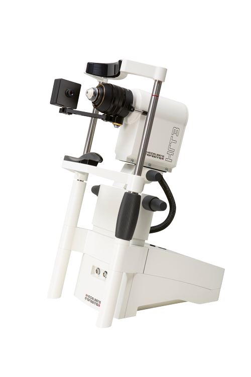
Heidelberg Engineering brings back in vivo corneal microscope | Heidelberg  Engineering GmbH
Heidelberg Engineering brings back in vivo corneal microscope | Heidelberg Engineering GmbH

Corneal confocal microscopy image acquisition part 1 - YouTube

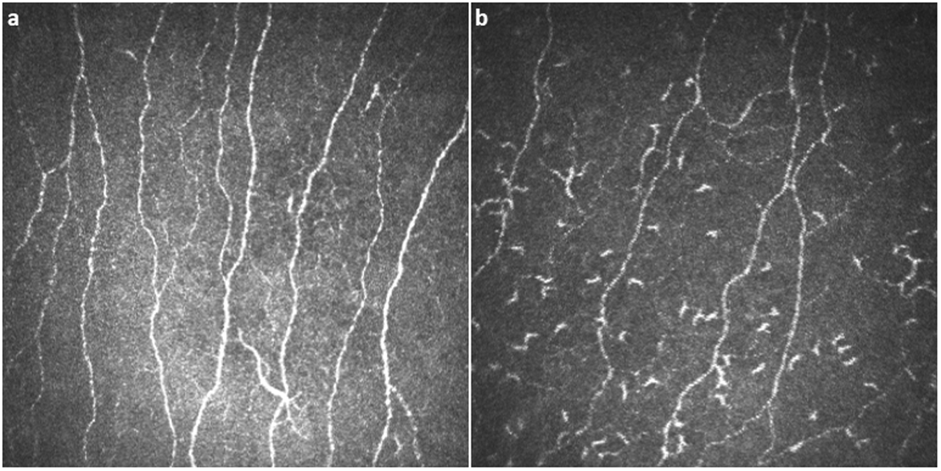
Corneal confocal microscopy detects corneal nerve damage and increased  dendritic cells in Fabry disease | Scientific Reports
Corneal confocal microscopy detects corneal nerve damage and increased dendritic cells in Fabry disease | Scientific Reports

Clinical in vivo confocal microscopy of the human cornea in health and  disease - ScienceDirect
Clinical in vivo confocal microscopy of the human cornea in health and disease - ScienceDirect

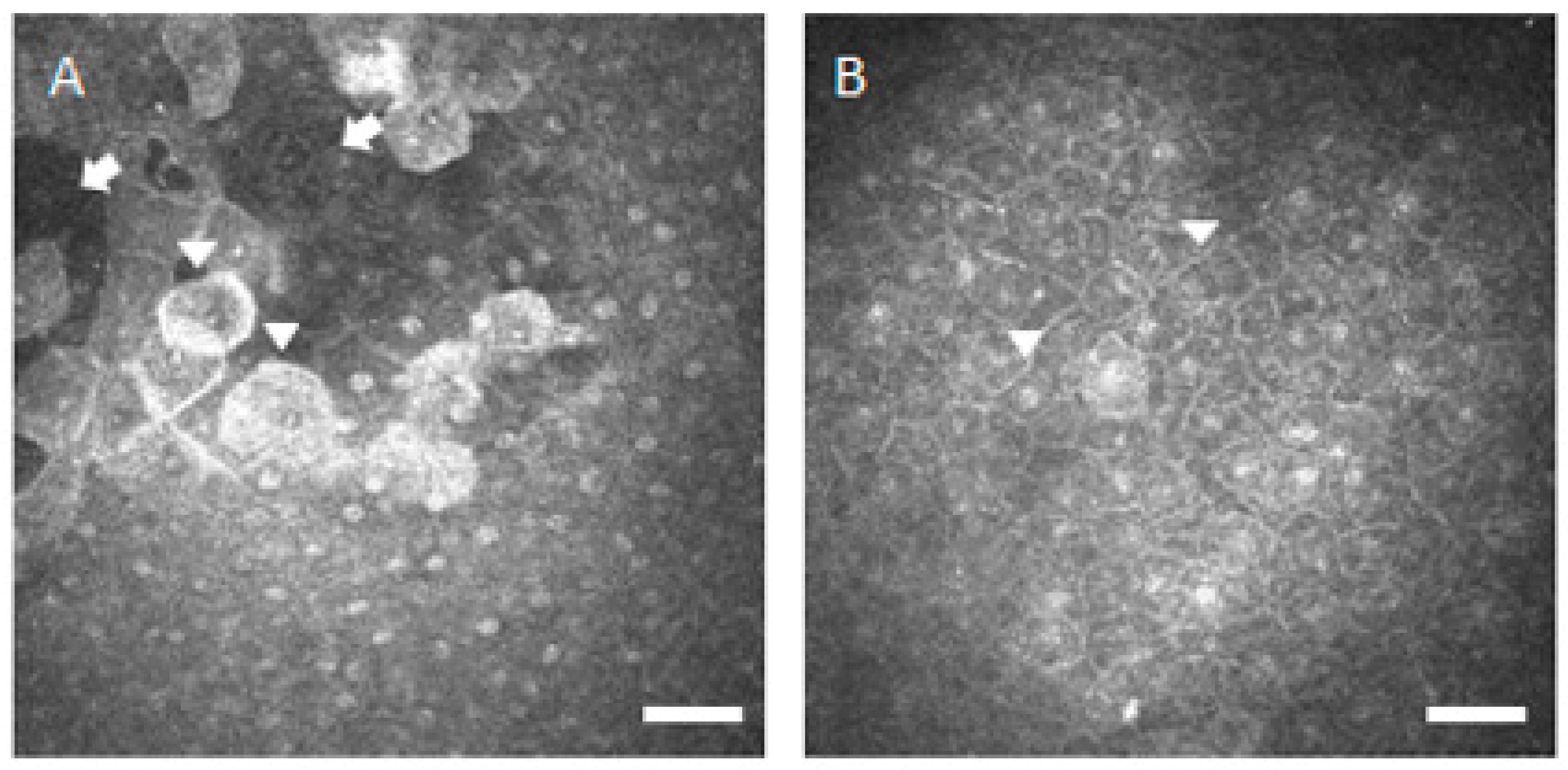
In vivo confocal microscopy of the corneal stroma and endothelium 24 h... |  Download Scientific Diagram
In vivo confocal microscopy of the corneal stroma and endothelium 24 h... | Download Scientific Diagram

3 Corneal confocal microscopy image of a control subject ( right panel... |  Download Scientific Diagram
3 Corneal confocal microscopy image of a control subject ( right panel... | Download Scientific Diagram

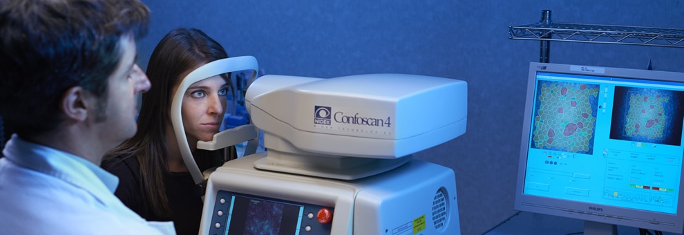
Corneal confocal microscopy (CONFOSCAN) - Centro Vista

In vivo confocal microscopy of the cornea and the limbus in a normal... |  Download Scientific Diagram
In vivo confocal microscopy of the cornea and the limbus in a normal... | Download Scientific Diagram

Pathophysiology | Free Full-Text | Corneal Confocal Microscopy in the  Diagnosis of Small Fiber Neuropathy: Faster, Easier, and More Efficient  Than Skin Biopsy?
Pathophysiology | Free Full-Text | Corneal Confocal Microscopy in the Diagnosis of Small Fiber Neuropathy: Faster, Easier, and More Efficient Than Skin Biopsy?
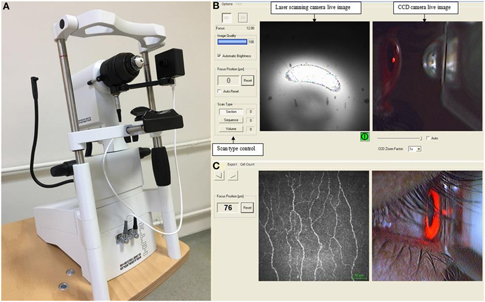
Frontiers | In Vivo Confocal Microscopic Evaluation of Corneal Nerve Fibers  and Dendritic Cells in Patients With Behçet's Disease
Frontiers | In Vivo Confocal Microscopic Evaluation of Corneal Nerve Fibers and Dendritic Cells in Patients With Behçet's Disease

CS4 Corneal Confocal Microscope - YouTube

Combining In Vivo Corneal Confocal Microscopy with Deep Learning-based  Analysis Reveals Sensory Nerve Fiber Loss in Acute SIV Infection | bioRxiv
Combining In Vivo Corneal Confocal Microscopy with Deep Learning-based Analysis Reveals Sensory Nerve Fiber Loss in Acute SIV Infection | bioRxiv
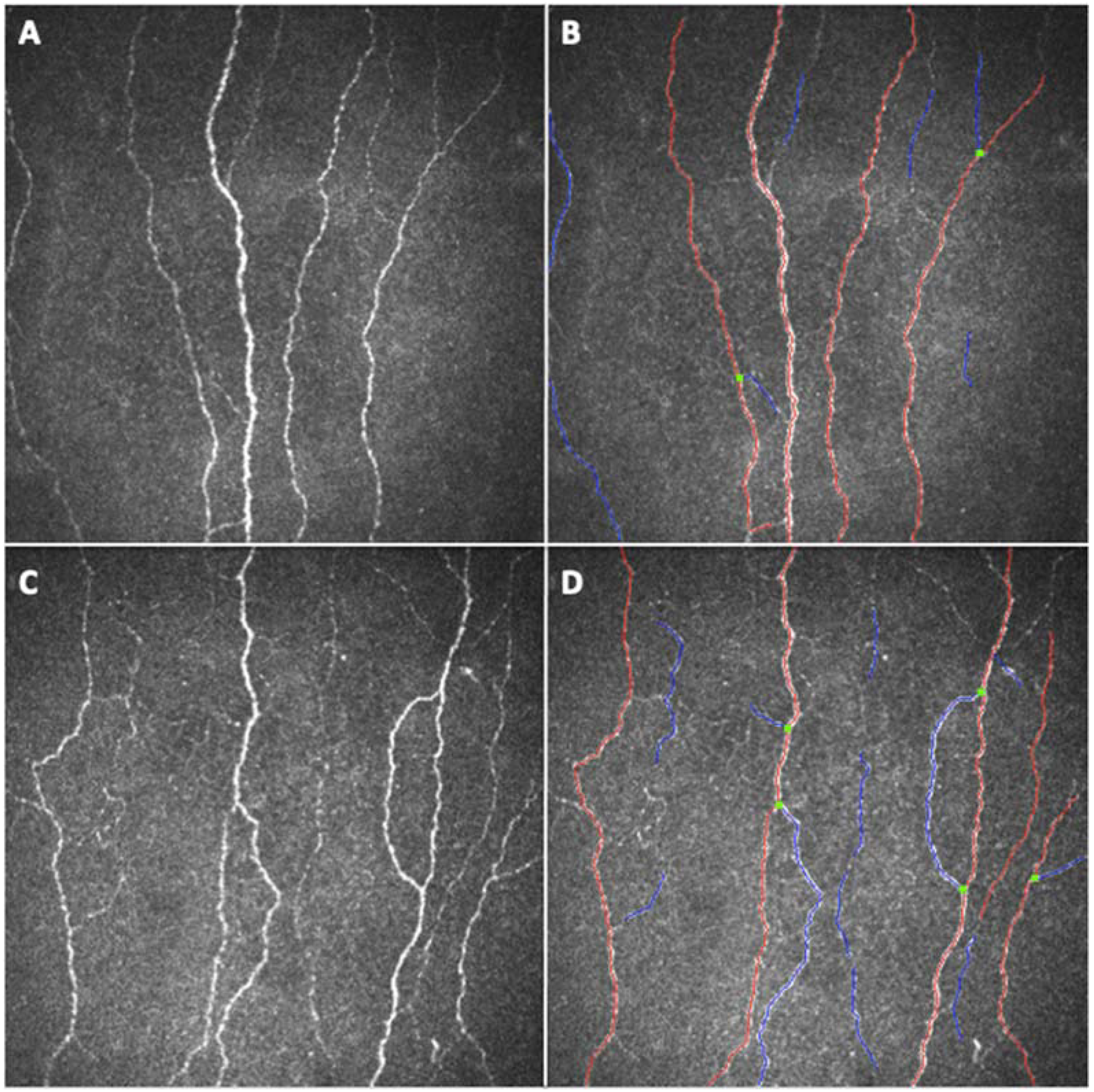
Confocal microscopy: Seeing what does not meet the eye

JPM | Free Full-Text | Small Fibre Peripheral Alterations Following  COVID-19 Detected by Corneal Confocal Microscopy
JPM | Free Full-Text | Small Fibre Peripheral Alterations Following COVID-19 Detected by Corneal Confocal Microscopy
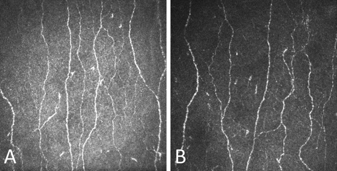
Corneal confocal microscopy detects small fibre neurodegeneration in  Parkinson's disease using automated analysis | Scientific Reports
Corneal confocal microscopy detects small fibre neurodegeneration in Parkinson's disease using automated analysis | Scientific Reports
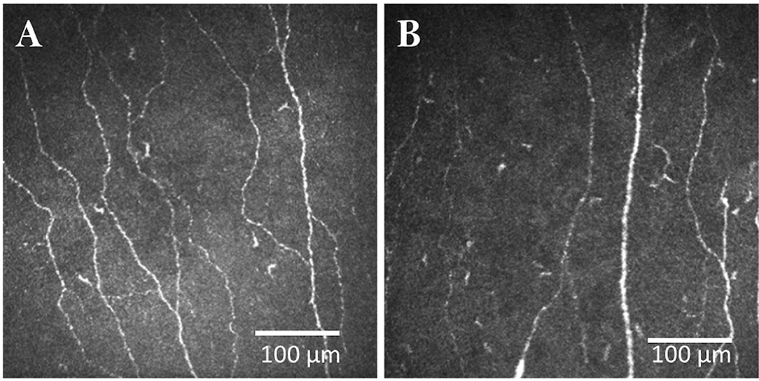
Frontiers | Corneal Confocal Microscopy to Image Small Nerve Fiber  Degeneration: Ophthalmology Meets Neurology
Frontiers | Corneal Confocal Microscopy to Image Small Nerve Fiber Degeneration: Ophthalmology Meets Neurology

Corneal confocal microscopy detects small nerve fibre damage in patients  with painful diabetic neuropathy | Scientific Reports
Corneal confocal microscopy detects small nerve fibre damage in patients with painful diabetic neuropathy | Scientific Reports

SciELO - Brasil - Corneal confocal microscopy in a healthy Brazilian sample Corneal  confocal microscopy in a healthy Brazilian sample
SciELO - Brasil - Corneal confocal microscopy in a healthy Brazilian sample Corneal confocal microscopy in a healthy Brazilian sample

Corneal confocal microscopy compared with quantitative sensory testing and  nerve conduction for diagnosing and stratifying the severity of diabetic  peripheral neuropathy | BMJ Open Diabetes Research & Care
Corneal confocal microscopy compared with quantitative sensory testing and nerve conduction for diagnosing and stratifying the severity of diabetic peripheral neuropathy | BMJ Open Diabetes Research & Care

One-Year Follow-up of Corneal Confocal Microscopy After Corneal  Cross-Linking in Patients With Post Laser In Situ Keratosmileusis Ectasia  and Keratoconus - American Journal of Ophthalmology
One-Year Follow-up of Corneal Confocal Microscopy After Corneal Cross-Linking in Patients With Post Laser In Situ Keratosmileusis Ectasia and Keratoconus - American Journal of Ophthalmology